Comparative Study of Macerated and Soxhlet-Extracted Moringa Oleifera Leaf Extracts: LC-MS-Based Metabolomic Profiling, Antioxidant Activity, and In Silico Target Prediction
DOI:
https://doi.org/10.22437/chp.v9i1.36870Keywords:
Antioxidant activity, In silico, metabolomic profiling, Moringa oleiferaAbstract
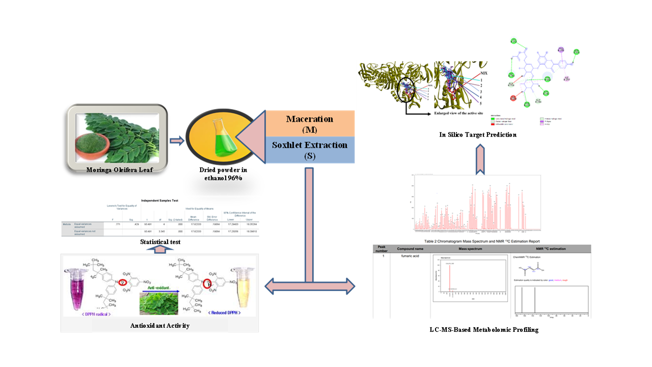
Moringa leaves (Moringa oleifera L.) are rich in secondary metabolites such as flavonoids, alkaloids, tannins, saponins, and terpenoids, which function as natural antioxidants. This study aimed to analyze the metabolite profile of M. oleifera leaf extracts obtained through two extraction techniques using LC-MS, evaluate their antioxidant activity via the DPPH assay, and predict the interaction between NADPH oxidase (as a receptor) and key plant-derived compounds through molecular docking. LC-MS results indicated that the maceration method yielded 101 secondary metabolites, with flavonoid derivatives comprising 70.99% of the extract, dominated by five key compounds including Kaempferol 3-O-robinobioside and Luteolin-7-glucoside. In contrast, the Soxhlet method resulted in 83 identified compounds, with a higher proportion of flavonoids (75.61%), and prominent compounds including quercetin-3-O-glucoside and Kaempferol 3-(6G-malonylneohesperidoside). Antioxidant testing with DPPH at concentrations of 10, 50, and 100 ppm revealed the Soxhlet extract had a stronger activity (IC₅₀ = 14.328 ppm) compared to the macerated extract (IC₅₀ = 32.092 ppm), with statistically significant differences (p < 0.05). Molecular docking demonstrated that Kaempferol 3-(6G-malonylneohesperidoside) exhibited the strongest binding affinity to NADPH oxidase (-10.1 kcal/mol), followed by other flavonoid derivatives. These findings underscore the antioxidant potential of M. oleifera, particularly from Soxhlet extraction, and suggest its promising application in pharmaceutical development as a natural antioxidant source.
Downloads
References
[1] Nofanda, Dwi IWI. Skrining Fitokimia Dan Pengaruh Metode Ekstraksi Terhadap Uji Aktivitas Antioksidan Ekstrak Etanol Daun Kelor (Moringa oleifera L.) dengan Metode DPPH. Diss. Universitas dr. SOEBANDI. 2022.
[2] Fakriah, E. Kurniasih, Adriana, and Rusydi. Sosialisasi Bahaya Radikal Bebas dan Fungsi Antioksidan Alami Bagi Kesehatan. J. Vokasi. 2019;3(1): 1-7. doi: 10.30811/vokasi.v3i1.960.
[3] Simanjuntak EJ, Zulham Z. Superoksida Dismutase (SOD) dan radikal bebas. Jurnal Keperawatan dan Fisioterapi (Jkf). 2020;2(2):124-129. doi: 10.35451/jkf.v2i2.342.
[4] Pham-Huy LA, He H, Pham-Huy C. Free radicals, antioxidants in disease and health. International journal of biomedical science: IJBS. 2008;4(2):89.
[5] Giovanni Martemucci, Ciro Costagliola, Michele Mariano , Luca D’andrea, Pasquale Napolitano and Angela Gabriella D’Alessandro. Free Radical Properties, Source and Targets, Antioxidant Consumption and Health. Oxygen. 2022;2:48-78. doi: https://doi.org/10.3390/oxygen2020006.
[6] Hadidi M, Orellana-Palacios JC, Aghababaei F, Gonzalez-Serrano DJ, Moreno A, Lorenzo JM. Plant by-product antioxidants: Control of protein-lipid oxidation in meat and meat products. Lwt. 2022;169:114003.
[7] Chukwuebuka E. Moringa oleifera “the mother’s best friend”. International Journal of Nutrition and Food Sciences. 2015;4(6):624-30. doi: 10.11648/j.ijnfs.20150406.14.
[8] Kamal SE, Aris M. Aktivitas antioksidan ekstrak etanol 70% daun kelor (Moringa oleifera Lam.) Terhadap DPPH. Jurnal Pro-Life. 2021;8(2):168-77.
[9] Sreelatha S, Padma PR. Antioxidant activity and total phenolic content of Moringa oleifera leaves in two stages of maturity. Plant foods for human nutrition. 2009;64:303-11. doi: 10.1007/s11130-009-0141-0.
[10] Faizal IA, Alifah AA. Perbandingan Metode Maserasi Dan Soxhletasi Ekstrak Daun Sirih Merah (Piper crocatum Ruiz & Pav) Terhadap Efektivitas Bakteri Staphylococcus epidermidis. Lumbung Farmasi: Jurnal Ilmu Kefarmasian. 2023;4(1):64-72. doi: 10.31764/lf.v4i1.10728.
[11] Meigaria KM, Mudianta IW, Martiningsih NW. Skrining fitokimia dan uji aktivitas antioksidan ekstrak aseton daun kelor (Moringa oleifera). Wahana Matematika dan Sains: Jurnal Matematika, Sains, dan Pembelajarannya. 2016;10(2):1-1. doi: https://doi.org/10.23887/wms.v10i2.12659.
[12] A. Saputra, F. Arfi, and M. Yulian. Literature Review: Analisis Fitokimia dan Manfaat Ekstrak Daun Kelor (Moringa oleifera). Amina. 2020;2(3): 114–119.
[13] Azalia D, Rachmawati I, Zahira S, Andriyani F, Sanini TM, Supriyatin S, Aulya NR. Uji Kualitatif Senyawa Aktif Flavonoid Dan Terpenoid Pada Beberapa Jenis Tumbuhan Fabaceae Dan Apocynaceae Di Kawasan Tngpp Bodogol. Bioma: Jurnal Biologi Makassar. 2023;8(1):32-43.
[14] Muthmainnah B. Skrining fitokimia senyawa metabolit sekunder dari ekstrak etanol buah delima (Punica granatum L.) dengan metode uji warna. Media Farmasi. 2017;13 (2): 23–28.
[15] Ikalinus R, Widyastuti SK, Setiasih NL. Skrining fitokimia ekstrak etanol kulit batang kelor (Moringa oleifera). Indonesia Medicus Veterinus. 2015;4(1):71-9.
[16] Marliana SD, Suryanti V, Suyono S. Skrining fitokimia dan analisis kromatografi lapis tipis komponen kimia buah labu siam (Sechium edule Jacq. Swartz.) dalam ekstrak etanol. Biofarmasi. 2005;3(1):26-31.
[17] Listiana L, Wahlanto P, Ramadhani SS, Ismail R. Penetapan Kadar Tanin Dalam Daun Mangkokan (Nothopanax scutellarium Merr) Perasan dan Rebusan Dengan Spektrofotometer UV-Vis. Pharmacy Genius. 2022;1(1):62-73. doi: 10.56359/pharmgen.v1i01.152.
[18] Fransiska AN, Masyrofah D, Marlian H, Sakina IV, Tyasna PS. Identifikasi senyawa terpenoid dan steroid pada beberapa tanaman menggunakan pelarut n-heksan. Jurnal Health Sains. 2021;2(6):733-41. DOI: 10.36387/Jiis.V4i1.285.
[19] Masadi YI, Lestari T, Dewi IK. Identifikasi Kualitatif Senyawa Terpenoid Ekstrak N-Heksana Sediaan Losion Daun Jeruk Purut (Citrus hystrix DC). Jurnal Kebidanan Dan Kesehatan Tradisional. 2018;3(1).
[20] R Kamal SE, Aris M. Aktivitas antioksidan ekstrak etanol 70% daun kelor (Moringa oleifera Lam.) Terhadap DPPH. Jurnal Pro-Life. 2021;8(2):168-77.
[21] Heim KE, Tagliaferro AR, Bobilya DJ. Flavonoid antioxidants: chemistry, metabolism and structure-activity relationships. The Journal of nutritional biochemistry. 2002;13(10):572-84.
[22] Chu YH, Chang CL, Hsu HF. Flavonoid content of several vegetables and their antioxidant activity. Journal of the Science of Food and Agriculture. 2000;80(5):561-6.
[23] Kumar S, Pandey AK. Chemistry and biological activities of flavonoids: an overview. The scientific world journal. 2013;2013(1):162750.
[24] Rosita JM, Taufiqurrahman I, Edyson E. Perbedaan Total Flavonoid Antara Metode Maserasi Dengan Sokletasi Pada Ekstrak Daun Binjai (Mangifera caesia)(Studi pendahuluan terhadap proses pembuatan sediaan obat penyembuhan luka). Dentin. 2019;1(1).
[25] Rudiana T, Nurbayti S, Ashari TH, Zhorif SA, Suryani N. Comparison of Maceration and Soxhletation Methods on the Antioxidant Activity of the Bouea macrophylla Griff Plant. Jurnal Kimia Valensi. 2023;9(2):244-52.
[26] Larasati RA, Utami N, Lindawati NY. The Effect of Maceration Method and Soxhletation Method on Total Phenolic Content and Antioxidant Activity of Kirinyuh (Chromolaena odorata (L.) RM King & H. Rob) LEAF. Medical Sains: Jurnal Ilmiah Kefarmasian. 2023;8(3):1207-14.
[27] Karisma DI, Indriasari C. The Effect of Maceration and Soxhletation Extraction Methods on The Flavonoid Content of Anting-anting Leaves Extracts (Acalypha indica L.) Using Uv-Vis Spectrophotometry. Strada Journal of Pharmacy. 2023;5(2):73-9.
[28] Biehn SE, Lindert S. Accurate protein structure prediction with hydroxyl radical protein footprinting data. Nature communications. 2021;12(1):341. doi:https://doi.org/10.1038/s41467-020-20549-7
[29] Muadifah A, Tilarso DP, Sowe MS, Tarigan IL, Ngibad K, Yuliantari SD. LC-MS Based Metabolite Profiling Leaves Extract of Pluchea indica With Antioxidant Activity. Chempublish Journal. 2024;8(2):75-89. doi: https://doi.org/10.22437/chp.v8i2.36869.
[30] Lountos GT, Jiang R, Wellborn WB, Thaler TL, Bommarius AS, Orville AM. The crystal structure of NAD(P)H oxidase from Lactobacillus sanfranciscensis: insights into the conversion of O2 into two water molecules by the flavoenzyme. Biochemistry. 2006;45(32):9648-59.
Published
Versions
- 2026-01-20 (2)
- 2025-06-01 (1)